
Use cases
OrientationJ in the literature
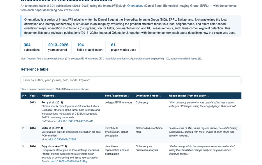
More than 300 peer-reviewed publications have used OrientationJ since 2013, across some two hundred fields: the actin cytoskeleton, collagen in tumors, electrospun nanofibers, engineered cardiac tissue, bone and mineralized tissue, and further afield geology, cryo-electron microscopy and liquid-crystal patterns. The reference table lists them all, each with the sentence from the paper describing how the plugin was used and which command it relied on. It sorts by year, field or mode, and it searches.
External resources
OrientationJ, the original page and OrientationPy — from the Biomedical Imaging Group.
Source code on GitHub — the Java source code under GPL-3.0 and the releases.
MonogenicJ — the companion plugin: wavelet-based multiresolution monogenic analysis (orientation, coherency, wavenumber per scale), bundled with OrientationJ since version 2.0.7.
OrientationPy on PyPI — pip install orientationpy, with a napari plugin.
image.sc forum — where questions on the plugins and their parameters are asked and answered; tag the post orientationj. Bugs and wishes go to the GitHub issues.
BIII, the BioImage Informatics Index — the curated registry entry: function, platform, licence and links, in the NEUBIAS tools index.
Comparisons, validation and other tools
Validation of structure tensor analysis — Gray B, Smith A, MacKenzie-Graham A, Shattuck D, Tward D (2025), Validation of structure tensor analysis for orientation estimation in brain tissue microscopy, Journal of Neuroscience Methods 423:110539. A simulation study of the method itself.
Comparison of fiber orientation analysis methods — a Thermo Fisher white paper comparing the usual approaches on the same images; vendor literature rather than a peer-reviewed study, but a compact overview of what each family of methods measures.
FiberFit — Morrill EE, Tulepbergenov AN, Stender CJ, Lamichhane R, Brown RJ, Lujan TJ (2016), A validated software application to measure fiber organization in soft tissue, Biomech Model Mechanobiol 15:1467–1478. Compares Fourier-based and structure-tensor fiber orientation; the validation reference listed on the original OrientationJ page.
DiameterJ — Hotaling NA, Bharti K, Kriel H, Simon CG (2015), DiameterJ: a validated open source nanofiber diameter measurement tool, Biomaterials 61:327–338. The fiber-diameter companion many nanofiber papers run alongside OrientationJ.
FiberO — Muñoz A, Docaj A, Fernandez J, Carriero A (2025), Front Bioeng Biotechnol. A recent open-source tool benchmarked against OrientationJ, Directionality, FiberFit and CT-FIRE.
Directionality (Fiji) — the Fourier-component and local-gradient alternative for orientation histograms, frequently used beside OrientationJ.
CT-FIRE — fiber tracking and extraction for collagen in second-harmonic-generation images.
skimage.feature.structure_tensor — the minimal structure tensor of scikit-image (finite-difference gradients only).